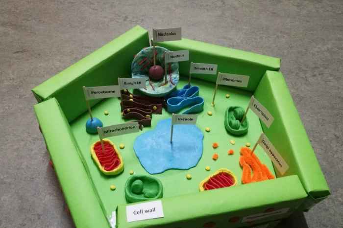
3D Cell Model Project is shaking up the world of biological research, offering a more realistic and insightful approach to studying cells and their functions. Gone are the days of flat, 2D cell cultures that only tell half the story.
These 3D models are revolutionizing how we understand disease, test drugs, and even develop personalized medicine.
Imagine tiny, self-organizing structures mimicking the complex environment of real organs, allowing scientists to see how cells interact, respond to treatments, and even develop diseases. This level of detail and complexity simply isn’t possible with traditional 2D models. This shift to 3D is leading to breakthroughs in drug discovery, personalized medicine, and regenerative medicine, making a real difference in human health.
Introduction to 3D Cell Models
3D cell models are revolutionizing biological research by providing a more realistic and physiologically relevant environment for studying cells and tissues. Unlike traditional 2D cell cultures, which are grown on flat surfaces, 3D cell models mimic the complex three-dimensional architecture and interactions found in the human body.
Advantages of 3D Cell Models
3D cell models offer several advantages over 2D cell cultures, making them a powerful tool for scientific investigations:
- Enhanced Cell-Cell and Cell-Matrix Interactions:3D models allow cells to interact with each other and with the surrounding extracellular matrix (ECM) in a more natural way, replicating the complex interactions found in vivo.
- Improved Cell Morphology and Function:Cells grown in 3D environments exhibit more realistic morphology and function, including cell differentiation, migration, and drug response.
- Greater Physiological Relevance:3D models provide a more accurate representation of the in vivo environment, leading to more reliable and clinically relevant results.
Types of 3D Cell Models
Several types of 3D cell models are available, each with its own advantages and limitations:
- Spheroids:Spheroids are aggregates of cells that spontaneously self-assemble into three-dimensional structures. They are relatively simple to create and can be used to study a variety of cellular processes.
- Organoids:Organoids are more complex 3D models that mimic the structure and function of specific organs. They are generated from stem cells or organ-specific cells and can be used to study organ development, disease pathogenesis, and drug response.
- Microfluidic Devices:Microfluidic devices are microchips that create controlled microenvironments for cell culture. They allow for precise control over cell culture conditions, such as nutrient delivery and waste removal, and can be used to study cell behavior in response to different stimuli.
Applications of 3D Cell Models
3D cell models have a wide range of applications in various fields, including drug discovery, toxicology testing, and disease modeling.
Drug Discovery and Development
3D cell models are increasingly used in drug discovery and development to assess drug efficacy and toxicity. They provide a more realistic platform for evaluating drug interactions with cells and tissues, compared to traditional 2D cell cultures.
- Drug Screening:3D cell models can be used to screen large libraries of compounds for potential drug candidates, identifying those with the most promising therapeutic effects.
- Toxicity Testing:3D models can help predict drug toxicity by assessing the effects of drugs on cells and tissues in a more physiologically relevant environment.
- Personalized Medicine:3D cell models derived from patient cells can be used to develop personalized treatments tailored to individual patients.
Toxicology Testing
3D cell models are being explored as alternatives to animal testing in toxicology studies. They provide a more humane and ethical approach to evaluating the safety of chemicals and drugs.
- Chemical Toxicity Assessment:3D models can be used to assess the toxic effects of chemicals on cells and tissues, providing insights into potential risks to human health.
- Environmental Toxicity:3D models can be used to study the effects of environmental pollutants on human cells and tissues, contributing to environmental protection efforts.
Disease Modeling
3D cell models are valuable tools for studying the mechanisms of disease and developing new therapies. They allow researchers to recreate the complex cellular and molecular events that occur in various diseases.
- Cancer Research:3D tumor models can be used to study tumor growth, invasion, and metastasis, and to evaluate the effectiveness of anticancer drugs.
- Neurodegenerative Diseases:3D brain organoids can be used to model neurodegenerative diseases such as Alzheimer’s disease and Parkinson’s disease, providing insights into disease progression and potential therapeutic targets.
- Diabetes Research:3D pancreatic islet models can be used to study the mechanisms of insulin production and secretion, and to evaluate potential treatments for diabetes.
Techniques for Creating 3D Cell Models
Several techniques are used to create 3D cell models, each with its own advantages and limitations.
Bioprinting
Bioprinting is a promising technique for creating complex 3D cell models by depositing cells and biomaterials in a layer-by-layer fashion. This method allows for precise control over cell placement and tissue architecture.
- Advantages:High precision, complex tissue structures, potential for organ regeneration.
- Limitations:Expensive, requires specialized equipment, limited scalability.
Microfluidic Devices
Microfluidic devices use microchannels to create controlled microenvironments for cell culture. They allow for precise control over cell culture conditions, such as nutrient delivery and waste removal.
- Advantages:Precise control over cell culture conditions, high throughput, potential for automated cell culture.
- Limitations:Requires specialized equipment, limited scalability, may not be suitable for all cell types.
Scaffold-Based Methods

Scaffold-based methods use biodegradable materials, such as hydrogels, to provide a structural support for cell growth. Cells can be seeded onto the scaffold and allowed to grow and interact with the surrounding material.
- Advantages:Relatively simple to create, can be used for a variety of cell types, allows for tissue regeneration.
- Limitations:Scaffold properties can influence cell behavior, may not be suitable for all cell types, can be challenging to control cell organization.
Characterization and Analysis of 3D Cell Models
Various methods are used to characterize and analyze 3D cell models, providing insights into cell behavior, function, and response to stimuli.
Microscopy
Microscopy is a fundamental technique for visualizing and analyzing 3D cell models. It allows researchers to study cell morphology, distribution, and interactions within the 3D environment.
- Confocal Microscopy:Provides high-resolution images of cells and tissues in 3D, allowing for detailed analysis of cell structure and function.
- Two-Photon Microscopy:Enables imaging of deeper tissue structures with reduced photodamage, making it suitable for studying thick 3D models.
Flow Cytometry
Flow cytometry is a technique for analyzing the properties of individual cells within a population. It can be used to assess cell viability, proliferation, and differentiation in 3D models.
- Cell Sorting:Flow cytometry can be used to isolate specific cell populations based on their properties, enabling further analysis.
- Multi-Parameter Analysis:Flow cytometry allows for simultaneous analysis of multiple cellular parameters, providing a comprehensive view of cell behavior.
Gene Expression Analysis, 3d cell model project
Gene expression analysis is used to study the changes in gene expression that occur in response to different stimuli or during disease progression. It provides insights into the molecular mechanisms underlying cell behavior in 3D models.
- RNA Sequencing:Provides a comprehensive view of gene expression patterns, identifying genes that are differentially expressed in 3D models.
- Quantitative PCR:Allows for the quantification of specific gene expression levels, providing insights into the regulation of specific genes.
Future Directions in 3D Cell Modeling
The field of 3D cell modeling is rapidly evolving, with exciting advancements in technology and techniques that promise to revolutionize biomedical research and healthcare.
Emerging Technologies
- Organ-on-a-Chip:Microfluidic devices that mimic the structure and function of specific organs, offering a more sophisticated platform for disease modeling and drug testing.
- Bioprinting with Advanced Materials:Development of new biomaterials and bioinks that mimic the properties of natural tissues, enabling the creation of more realistic and functional 3D models.
- Artificial Intelligence and Machine Learning:Application of AI and ML algorithms to analyze large datasets generated from 3D cell models, providing insights into complex biological processes.
Future Impact
3D cell models have the potential to transform biomedical research and healthcare by:
- Accelerating Drug Discovery and Development:Providing more relevant and reliable models for drug screening and toxicity testing, leading to faster and more efficient development of new therapies.
- Improving Disease Modeling and Understanding:Providing a more realistic platform for studying the mechanisms of disease, leading to the development of targeted therapies and personalized medicine.
- Reducing Animal Testing:Providing a more humane and ethical alternative to animal testing in toxicology and drug development.
Ending Remarks: 3d Cell Model Project
The future of 3D cell models is bright, promising a deeper understanding of human biology and leading to more effective treatments for a wide range of diseases. As we continue to refine these models and explore new applications, the potential for impact on human health is limitless.
It’s a fascinating field that’s pushing the boundaries of science and giving us a glimpse into a future where personalized medicine and innovative therapies are the norm.
FAQs
What are the main advantages of using 3D cell models?
3D cell models offer a more realistic representation of the cellular environment, allowing for more accurate studies of cell behavior, drug interactions, and disease progression. They also provide a more complex and dynamic environment for cells to grow and interact, mimicking the in vivo conditions more closely.
How do 3D cell models differ from traditional 2D cell cultures?
Unlike traditional 2D cell cultures, which are grown on a flat surface, 3D cell models mimic the three-dimensional structure and complexity of real tissues. This allows for more accurate studies of cell-cell interactions, signaling pathways, and tissue-specific responses.
What are some examples of diseases that can be studied using 3D cell models?
3D cell models have been successfully used to study a wide range of diseases, including cancer, Alzheimer’s disease, diabetes, and cardiovascular disease. These models allow researchers to study disease mechanisms, test potential therapies, and develop personalized treatments.


Leave a Comment