Functions of different parts of the brain – Unraveling the intricate functions of different brain regions, this article delves into the fascinating world of neurology, revealing the remarkable roles played by each part in shaping our thoughts, actions, and very existence.
From the cerebral cortex, responsible for higher-order cognitive functions, to the cerebellum, coordinating movement and balance, each brain region contributes uniquely to the symphony of our being.
Functions of the Cerebral Cortex
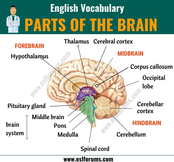
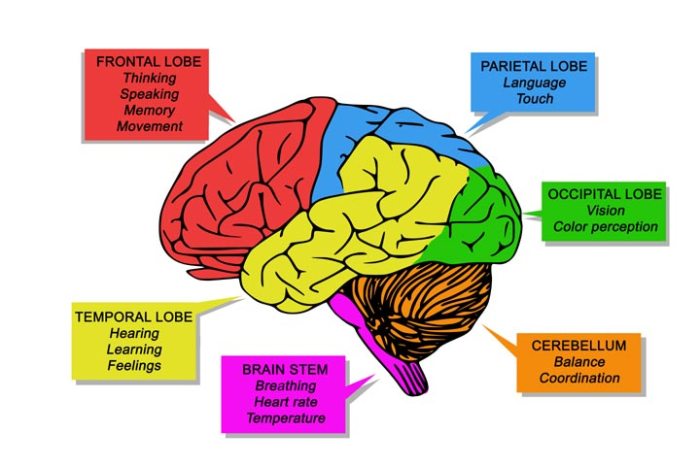
The cerebral cortex, the outermost layer of the brain, is responsible for higher-order cognitive functions such as language, memory, and decision-making. It is divided into two hemispheres, each of which is further divided into four lobes: the frontal lobe, parietal lobe, temporal lobe, and occipital lobe.
The cerebral cortex is highly specialized, with different regions responsible for different functions. The motor cortex, located in the frontal lobe, controls voluntary movement. The sensory cortex, located in the parietal lobe, receives and processes sensory information from the body.
The association areas, located in the temporal and occipital lobes, are involved in higher-order cognitive functions such as language, memory, and decision-making.
Motor Cortex
- Controls voluntary movement
- Located in the frontal lobe
- Different areas of the motor cortex control different body parts
Sensory Cortex
- Receives and processes sensory information from the body
- Located in the parietal lobe
- Different areas of the sensory cortex process different types of sensory information, such as touch, temperature, and pain
Association Areas
- Involved in higher-order cognitive functions such as language, memory, and decision-making
- Located in the temporal and occipital lobes
- Different areas of the association areas are responsible for different cognitive functions
Functions of the Basal Ganglia
The basal ganglia are a group of subcortical brain structures that play a crucial role in motor control, habit formation, and reward processing. They interact with other brain structures, such as the cerebral cortex and thalamus, to coordinate movement, learn new skills, and regulate motivation.
Motor Control
- The basal ganglia are involved in the initiation, execution, and termination of movement. They receive input from the cerebral cortex and thalamus and send output signals to the brainstem and spinal cord.
- The basal ganglia help to control the speed, force, and direction of movement. They also help to suppress unwanted movements, such as tics and tremors.
Habit Formation
- The basal ganglia are involved in the formation of habits. When we repeat a behavior over and over again, the basal ganglia create a neural pathway that makes it easier to perform that behavior in the future.
- This process is known as procedural memory. It allows us to learn new skills and habits without having to think about each step consciously.
Reward Processing
- The basal ganglia are involved in reward processing. They receive input from the reward system of the brain and help to determine whether a behavior is rewarding or not.
- This information is used to guide our behavior and to motivate us to seek out rewarding experiences.
Functions of the Thalamus
The thalamus is a small structure located deep within the brain, just above the brainstem. It serves as a relay center for sensory information, playing a crucial role in consciousness and sleep-wake cycles.The thalamus receives sensory information from all parts of the body except the olfactory bulb.
It then processes this information and sends it to the appropriate areas of the cerebral cortex for further processing. The thalamus also plays a role in motor control, memory, and emotion.
Nuclei of the Thalamus
The thalamus is divided into several nuclei, each with its own specific function. Some of the most important nuclei include:
Anterior nucleus
Receives sensory information from the body and sends it to the somatosensory cortex.
Ventral posterior nucleus
Receives sensory information from the face and sends it to the somatosensory cortex.
Lateral geniculate nucleus
Receives visual information from the retina and sends it to the visual cortex.
Medial geniculate nucleus
Receives auditory information from the cochlea and sends it to the auditory cortex.
Pulvinar nucleus
Receives sensory information from all parts of the body and sends it to the association cortex.
Functions of the Hypothalamus
The hypothalamus is a small region located at the base of the brain that plays a crucial role in regulating various bodily functions. It serves as a link between the nervous system and the endocrine system, influencing hormones and stress responses.
Homeostasis Regulation
The hypothalamus is responsible for maintaining homeostasis, the body’s internal balance. It monitors and adjusts body temperature, hunger, thirst, and sleep cycles to ensure optimal functioning. The hypothalamus detects changes in the body’s internal environment and initiates appropriate responses to maintain equilibrium.
Endocrine System Involvement, Functions of different parts of the brain
The hypothalamus is closely connected to the pituitary gland, which is often referred to as the “master gland” of the endocrine system. The hypothalamus produces releasing and inhibiting hormones that regulate the secretion of hormones from the pituitary gland. These hormones, in turn, influence the activity of other glands throughout the body, controlling various physiological processes.
Stress Responses
The hypothalamus is involved in the body’s stress response system. When the body experiences stress, the hypothalamus activates the sympathetic nervous system and the adrenal glands, releasing hormones such as adrenaline and cortisol. These hormones prepare the body for a “fight or flight” response, increasing heart rate, blood pressure, and energy levels.
Functions of the Cerebellum
The cerebellum is a vital part of the brain responsible for motor coordination, balance, and spatial navigation. It receives sensory information from the body and sends signals to the muscles to control movement and maintain equilibrium.
Parts of the Cerebellum
- Cerebellar Cortex:The outermost layer of the cerebellum, responsible for receiving sensory information and processing motor commands.
- Cerebellar Nuclei:Deep structures that relay information from the cerebellar cortex to other brain regions.
- White Matter:Connects different parts of the cerebellum and transmits signals to and from other brain areas.
Functions of the Cerebellum
- Motor Coordination:The cerebellum helps to coordinate muscle movements, ensuring smooth and precise execution of voluntary actions.
- Balance:It processes sensory information from the vestibular system to maintain balance and prevent falling.
- Spatial Navigation:The cerebellum assists in spatial orientation, allowing individuals to navigate their environment effectively.
Functions of the Brainstem
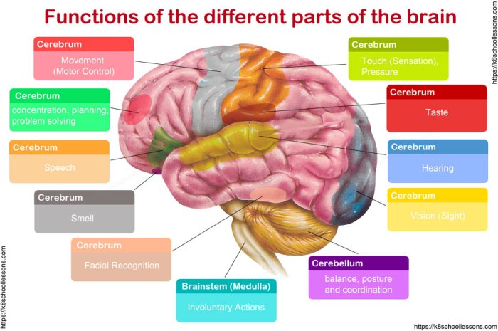
The brainstem is a vital part of the central nervous system that connects the cerebrum and cerebellum to the spinal cord. It is responsible for controlling many essential life functions, including breathing, heart rate, and blood pressure regulation.The brainstem is divided into three main parts: the medulla, pons, and midbrain.
Each part has its own specific functions.
Medulla
The medulla is the lowest part of the brainstem and is responsible for controlling vital life functions such as breathing, heart rate, and blood pressure. It also contains the nuclei of the cranial nerves that control swallowing, salivation, and tongue movement.
Pons
The pons is located above the medulla and is responsible for relaying sensory information from the spinal cord to the cerebrum. It also contains the nuclei of the cranial nerves that control facial expression, hearing, and balance.
Midbrain
The midbrain is the uppermost part of the brainstem and is responsible for controlling eye movements, pupillary reflexes, and motor coordination. It also contains the substantia nigra, which is a brain region that is involved in the production of dopamine, a neurotransmitter that is essential for motor control.
Functions of the Limbic System: Functions Of Different Parts Of The Brain
The limbic system is a complex network of brain structures located deep within the cerebrum. It plays a crucial role in our emotional experiences, memory formation, and motivation.The limbic system is made up of several key structures, including the amygdala, hippocampus, and cingulate cortex.
Each of these structures has a specific role in the limbic system’s functions.
Amygdala
The amygdala is a small almond-shaped structure located on either side of the brain. It is responsible for processing emotions, particularly fear and anxiety. The amygdala helps us to identify and respond to potential threats in our environment.
Hippocampus
The hippocampus is a curved structure located in the medial temporal lobe of the brain. It is essential for memory formation and consolidation. The hippocampus helps us to encode new memories and to retrieve them when we need them.
Cingulate Cortex
The cingulate cortex is a thin strip of tissue that encircles the corpus callosum. It is involved in a variety of functions, including emotion, motivation, and decision-making. The cingulate cortex helps us to weigh the pros and cons of different choices and to make decisions that are in our best interests.
Functional Connectivity of Brain Regions
The brain is a complex organ that is responsible for a wide range of functions, from movement and sensation to cognition and emotion. These functions are not carried out by individual brain regions working in isolation, but rather by the coordinated activity of multiple brain regions.
This coordination is known as functional connectivity.
Functional connectivity can be studied using a variety of techniques, including fMRI and EEG. fMRI measures changes in blood flow in the brain, which is an indirect measure of neural activity. EEG measures electrical activity in the brain directly.
Studies using these techniques have shown that the brain is organized into a number of functional networks. These networks are involved in a variety of different functions, such as attention, memory, and language.
The study of functional connectivity is still in its early stages, but it has the potential to provide us with a much better understanding of how the brain works.
Studying Functional Connectivity
There are a number of different techniques that can be used to study functional connectivity. One common technique is fMRI. fMRI measures changes in blood flow in the brain, which is an indirect measure of neural activity. When a brain region is active, it requires more blood flow.
fMRI can be used to measure changes in blood flow in different brain regions over time, and this information can be used to create a map of functional connectivity.
Another common technique for studying functional connectivity is EEG. EEG measures electrical activity in the brain directly. EEG can be used to measure the synchronization of electrical activity between different brain regions. This information can be used to create a map of functional connectivity that is similar to the one that can be created using fMRI.
Functional Connectivity and Brain Function
The study of functional connectivity has helped us to understand how the brain is organized into functional networks. These networks are involved in a variety of different functions, such as attention, memory, and language.
For example, the default mode network is a network of brain regions that is active when the brain is at rest. This network is involved in self-referential processing, such as thinking about oneself and one’s experiences.
The salience network is a network of brain regions that is involved in detecting and responding to salient stimuli. This network is active when we are paying attention to something or when we are surprised.
The central executive network is a network of brain regions that is involved in executive function, such as planning, decision-making, and working memory. This network is active when we are engaged in complex cognitive tasks.
These are just a few examples of the many functional networks that have been identified in the brain. The study of functional connectivity is still in its early stages, but it has the potential to provide us with a much better understanding of how the brain works.
Epilogue
In conclusion, the functions of different brain regions form a complex and awe-inspiring tapestry, underscoring the incredible capabilities of the human mind. As we continue to explore the intricacies of this remarkable organ, we unlock the potential for groundbreaking discoveries that will redefine our understanding of ourselves and our place in the world.
FAQ Summary
What is the role of the hypothalamus in the body?
The hypothalamus plays a crucial role in maintaining homeostasis, regulating body temperature, hunger, thirst, and sleep-wake cycles.
How does the cerebellum contribute to movement?
The cerebellum coordinates motor movements, ensuring precision, balance, and spatial navigation.
What is the function of the limbic system?
The limbic system is involved in processing emotions, forming memories, and motivating behavior.



Leave a Comment